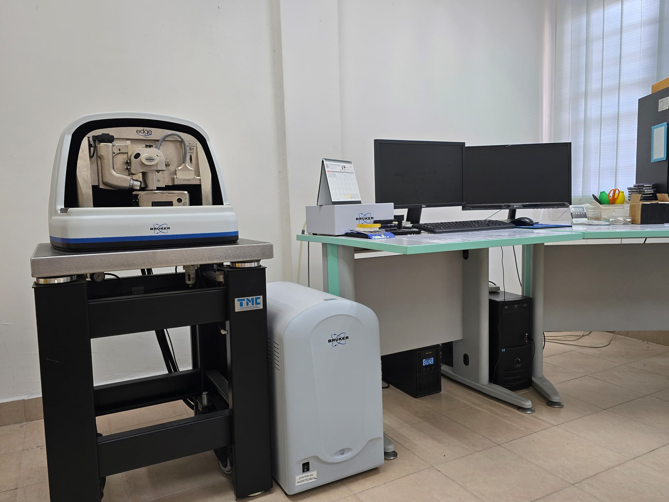
Brand name: Brunker Crest
Model name: Dimension Edge with Scanasyst
Description:
The Atomic force microscope allows us to investigate the structure and morphology of the surface. This is a powerful technique which generates topographic images of the structure with atomic resolution.
AFM used as an surface imaging tool:
- Scan the surface materials to produce topographical images
- Used to determine roughness, grain size, and features on the nanoscale
- Can resolve individual holes, defects and atomic clusters
AFM has been used for measurements on a wide variety of sample types such as thin and thick film coatings, ceramics, composites, glasses, synthetic and biological membrane, metals, polymers and semiconductors.
Scan available: 0-40 microns , 0-100 microns
Sample size: minimum 1 cm X 1 cm
Scan Mode :
- Scanasyst/Tapping/Contact
- Conductive AFM (1 DC bias)
- Magnetic Force Microscopy
- Piezoresponse Force Microscopy
- Force Spectroscopy
Location: Room 049 & 050, Block D, Faculty of Science 2, Universiti Putra Malaysia
Contact person:
 |
|
Nurul Shahida Ramli (Miss)
Assistant Science Officer
Tel: +603 97693233
|
| |
|
|
 |
|
Zulaikha Haziqah Mohd Zulkifli (Miss)
Assistant Science Officer
Tel: +603 97696656
|
Atomic Force Microscope (AFM) Form
BR045 Form
Updated:: 20/11/2024 [harithdaniel]
MEDIA SHARING